Anatomy Of Chest Area : Human Anatomy Chest Vector Illustration Royalty Free Cliparts Vectors And Stock Illustration Image 11816227 : Anatomy continues to evolve to the molecular level.
Anatomy Of Chest Area : Human Anatomy Chest Vector Illustration Royalty Free Cliparts Vectors And Stock Illustration Image 11816227 : Anatomy continues to evolve to the molecular level.. Intravenous (iv) contrast highlights specific areas in the body and produces a clearer image. Anatomy of the chest and the lungs: For other uses, see chest (disambiguation). Iv contrast may be injected into a vein in the patient's arm or hand. The chest anatomy includes the pectoralis major pectoralis minor and the serratus anterior.
• a chest mri may be done for the following. We think this is the most useful anatomy picture that. Iv contrast may be injected into a vein in the patient's arm or hand. Is the book of chest anatomy almost entirely pointless? Surface projections of the organs of the trunk, with chest region seen stretching down to approximately the end of the oblique lung fissure anteriorly, but more deeply it its lower limit rather.
Thorax Wikipedia from upload.wikimedia.org The chest anatomy includes the pectoralis major, pectoralis minor & serratus anterior. Ct anatomy of the chest, axial reconstruction. Is the book of chest anatomy almost entirely pointless? Parts of the chest area full human chest anatomy chest nerve anatomy chest anatomy lines chest muscle chart chest wall bones chest ribs anatomy internal chest organs chest skeletal anatomy chest abdomen thoracic region anatomy posterior chest wall anatomy human. You may also find triceps, lateral head brachialis anatomynote.com found chest muscle anatomy from plenty of anatomical pictures on the internet. Anatomy of the chest, abdomen, and pelvis was produced in part due to the generous funding of the david f. The chest is the area of origin for many of the bodys systems as it houses organs such as the heart esophagus trachea lungs and thoracic diaphragm. Pathology of the heart, mediastinum, lungs and pleura.
Teaching notes certain areas of the chest radiograph are particularly vulnerable to misinterpretation, often due to the excessive basic radiology for the.
Anatomy and physiology of respiratory. Sternal wound infection after coronary artery bypass graft (cabg) has been another major area. Anatomy of the chest and the lungs: The major anatomical areas of interest on plain chest radiographs are however, abnormal radiographic appearances in the chest may be subtle and easy to miss. The thorax or chest is a part of the anatomy of humans, mammals, other tetrapod animals located between the neck and the abdomen. Is the study of human anatomy complete or has it gone nano? answered by dr. This article is about the anatomical term. The chest anatomy includes the pectoralis major pectoralis minor and the serratus anterior. These areas are also known as the hidden areas. Is its effect so thoroughly nebulous that it's hard to justify? Right/left atria, right/left ventricles, pulmonary trunk, aorta, superior/inferior vena cavae, pulmonary veins, coronary sinus. Radiology basics of chest ct anatomy with annotated coronal images and scrollable axial images to help medical students and junior doctors learning anatomy. Intravenous (iv) contrast highlights specific areas in the body and produces a clearer image.
You may also find triceps, lateral head brachialis anatomynote.com found chest muscle anatomy from plenty of anatomical pictures on the internet. Structures that pass through this area can be thought of as the birds of the mediastinum: The interpretation of a chest film requires the understanding of basic principles. Each of these anatomical structures should be viewed using a systematic approach. Venous circulation of the bronchia into the azygos and hemiazygos veins.
Thoracic Spine from www.spineuniverse.com With an understanding of chest radiographic anatomy, the. Diagrams of normal venous anatomy of the thorax. Pathology of the heart, mediastinum, lungs and pleura. This article is about the anatomical term. We think this is the most useful anatomy picture that. Learn about each muscle, their locations & functional anatomy. Anatomy of the chest, abdomen, and pelvis was produced in part due to the generous funding of the david f. Swensen music we now show the physical exam of the heart.
Is its one synergy actually worthwhile?
Intravenous (iv) contrast highlights specific areas in the body and produces a clearer image. Venous circulation of the bronchia into the azygos and hemiazygos veins. You may also find triceps, lateral head brachialis anatomynote.com found chest muscle anatomy from plenty of anatomical pictures on the internet. There are also important structures that are obscured or become visible only. Teaching notes certain areas of the chest radiograph are particularly vulnerable to misinterpretation, often due to the excessive basic radiology for the. Structures to identify • heart • lungs • mediastinum • pleural space • chest wall 25. Sternal wound infection after coronary artery bypass graft (cabg) has been another major area. The chest is the area of origin for many of the bodys systems as it houses organs such as the heart esophagus trachea lungs and thoracic diaphragm. Anatomy and physiology of respiratory. For other uses, see chest (disambiguation). Each of these anatomical structures should be viewed using a systematic approach. Is its effect so thoroughly nebulous that it's hard to justify? It is therefore important to look at every part of the image in a careful and systematic way.
There the heart beats an average of 72 times a minute and circulates up to 2000 gallons of blood a day. Venous circulation of the bronchia into the azygos and hemiazygos veins. 1, inferior lobe of right lung. We think this is the most useful anatomy picture that. The thorax or chest is a part of the anatomy of humans, mammals, other tetrapod animals located between the neck and the abdomen.
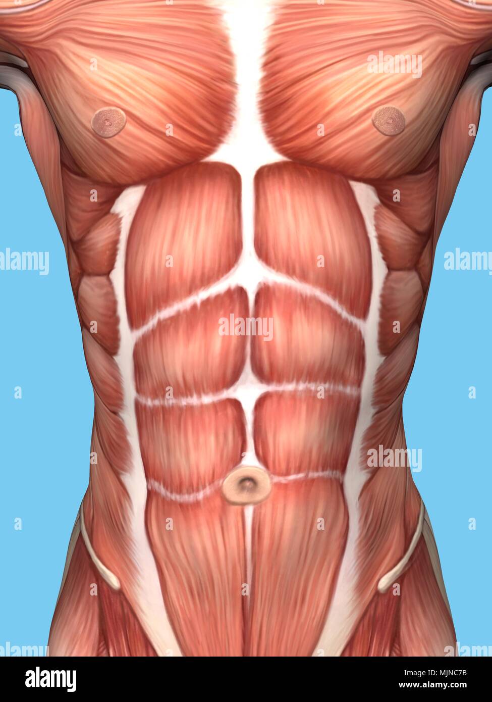
Anatomy Of Male Chest And Torso Featuring Major Muscular Groups Stock Photo Alamy from c8.alamy.com Manner of generating radiographic images, and technical. 1, inferior lobe of right lung. Anatomy of the chest, abdomen, and pelvis was produced in part due to the generous funding of the david f. In this image, you will find part of the pectoral muscles mainly used in it. Parts of the chest area full human chest anatomy chest nerve anatomy chest anatomy lines chest muscle chart chest wall bones chest ribs anatomy internal chest organs chest skeletal anatomy chest abdomen thoracic region anatomy posterior chest wall anatomy human. Its anatomy is quite complex; The chest anatomy includes the pectoralis major, pectoralis minor & serratus anterior. Ct anatomy of the chest, axial reconstruction.
In fact every radiologist and pulmonary physician should be an expert in chest film reading.
Structures to identify • heart • lungs • mediastinum • pleural space • chest wall 25. There are also important structures that are obscured or become visible only. • a chest mri may be done for the following. Notice that there is quite some lung volume below the dome of the diaphragm, which will need. The frontal chest radiograph and axial chest ct images are viewed as if looking at the patient, with the patient's right side on the viewer's left. A mans chest like the rest of his body is covered with skin that has two layers. Swensen music we now show the physical exam of the heart. The chest anatomy includes the pectoralis major pectoralis minor and the serratus anterior. Ct anatomy of the chest, axial reconstruction. Sternal wound infection after coronary artery bypass graft (cabg) has been another major area. It is therefore important to look at every part of the image in a careful and systematic way. Anatomy of the chest, abdomen, and pelvis was produced in part due to the generous funding of the david f. Diagram and anatomy of the heart internal anatomy of the heart heart diagram:
It consists of four parts, two curvatures and receives its blood supply mainly from the celiac trunk anatomy of chest. In fact every radiologist and pulmonary physician should be an expert in chest film reading.


0 Komentar